 Comptes Rendus Palevol
9 (6-7) - Pages 389-395
Comptes Rendus Palevol
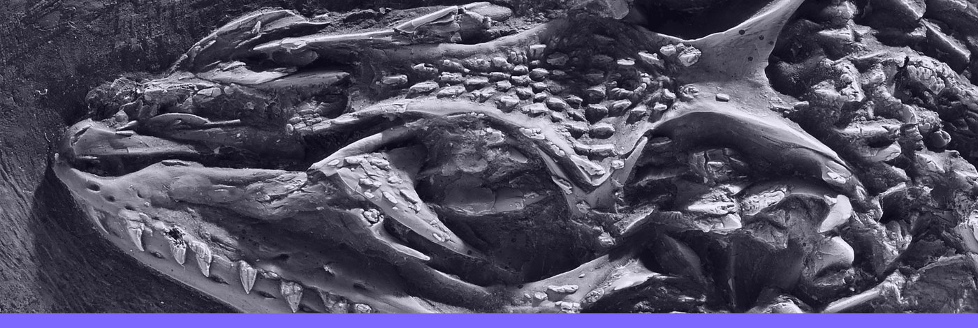
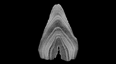
9 (6-7) - Pages 389-395Vertebrate microremains, particularly teeth, represent a substantial part of known vertebrate biodiversity. Many groups, such as Mesozoic mammals, are known mostly through isolated teeth. Classical imaging techniques of such complex millimetric to inframillime-tric objects are most often limited by problems of manipulation, depth of focus or limited orientation. The methods generally used are stereomicroscopy (including in-focus z-series reconstruction) and Scanning Electron Microscopy (SEM), which provide good images. Nevertheless, both provide 2D static images or partial and directional 3D data, making complete observation difficult. Propagation phase contrast synchrotron X-ray microtomography is a powerful technique alleviating these limitations. Thanks to submicron resolution and to the edge detection effect, it rapidly provides 3D data from minute samples with levels of quality and detail unattainable using conventional microtomographs. Complex morphology of small specimens can be studied with unlimited orientation possibilities and, when coupled with 3D printing, it allows enlarged 3D reproductions of such small and fragile fossils.
Microrestes, Imagerie, Synchrotron, Microtomographie, 3D, Contraste de phase