 Comptes Rendus Palevol
15 (8) - Pages 968-977
Comptes Rendus Palevol
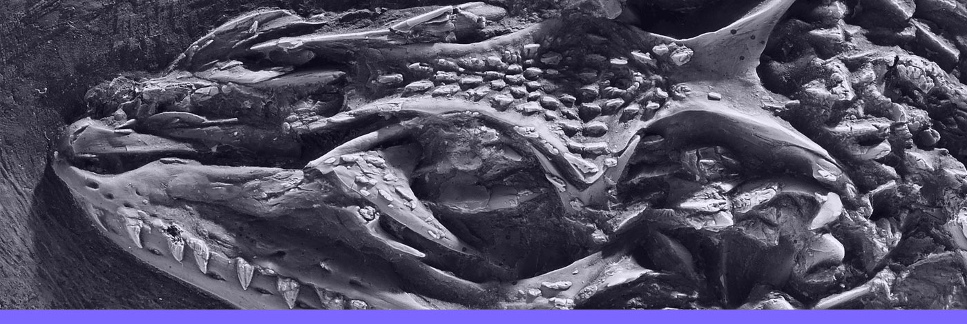
15 (8) - Pages 968-977Since the late 20th century, new technologies have provided powerful ways to digitize biological structures in three dimensions (3D). Among those, photogrammetry is a low cost and non-destructive method, which has become increasingly used since the development of the digital camera. Recent studies have demonstrated that reconstructions of isolated elements can be of as high quality as those obtained with laser scanners. Here, we wanted to test the performance of photogrammetry for the quantitative analysis of mounted specimens in museum exhibitions. Indeed, access to material can be an issue in comparative anatomy and, especially, in paleontology. This is notably the case for large, impressive specimens. We performed reconstructions based on acquisitions done under various conditions and also tested the reconstruction performance of two software programs. The resulting 3D models were then compared to a reference object corresponding to the bone of interest digitized with a cutting-edge surface scanner. Our results show that photogrammetry enables quality reconstruction of the almost entire surface of the mounted bone of interest. Photogrammetry thus appears a reliable method perfectly suited to study large specimens exposed in museum gallery.
Photogrammetry, Mounted skeletons, Paleontology, 3D, Digitization, Agisoft photoscan, VisualSFM
DOI (Digital Object Identifier)
https://doi.org/10.1016/j.crpv.2016.08.003
Permalink (to link this page)
http://cr-palevol.fr/15/95