
 Geodiversitas
31 (4) - Pages 851-863
Geodiversitas
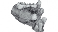
31 (4) - Pages 851-863Dental enamel thickness is commonly listed among the diagnostic features for taxonomic assessment and phylogenetic reconstruction in the study of fossil hominids, and is widely used as an indicator of dietary habits and palaeoenvironmental conditions. However, little quantitative information is currently available on its topographic variation in deciduous crowns of fossil primates. By means of high-resolution microtomography, we investigated the inner structural morphology of the mixed lower dentition of Ouranopithecus macedoniensis, a late Miocene large-bodied ape from Macedonia, Greece. With respect to the extant African apes and Homo, O. macedoniensis shows a significant difference in occlusal enamel thickness between the relatively thin deciduous second molar and the absolutely thick-enamelled permanent first molar.
Mammalia, Primates, Ouranopithecus, hominid, Miocene ape, mixed dentition, microtomography, tooth inner structure, 3D reconstruction, enamel thickness.