 Comptes Rendus Palevol
22 (32) - Pages 635-665
Comptes Rendus Palevol
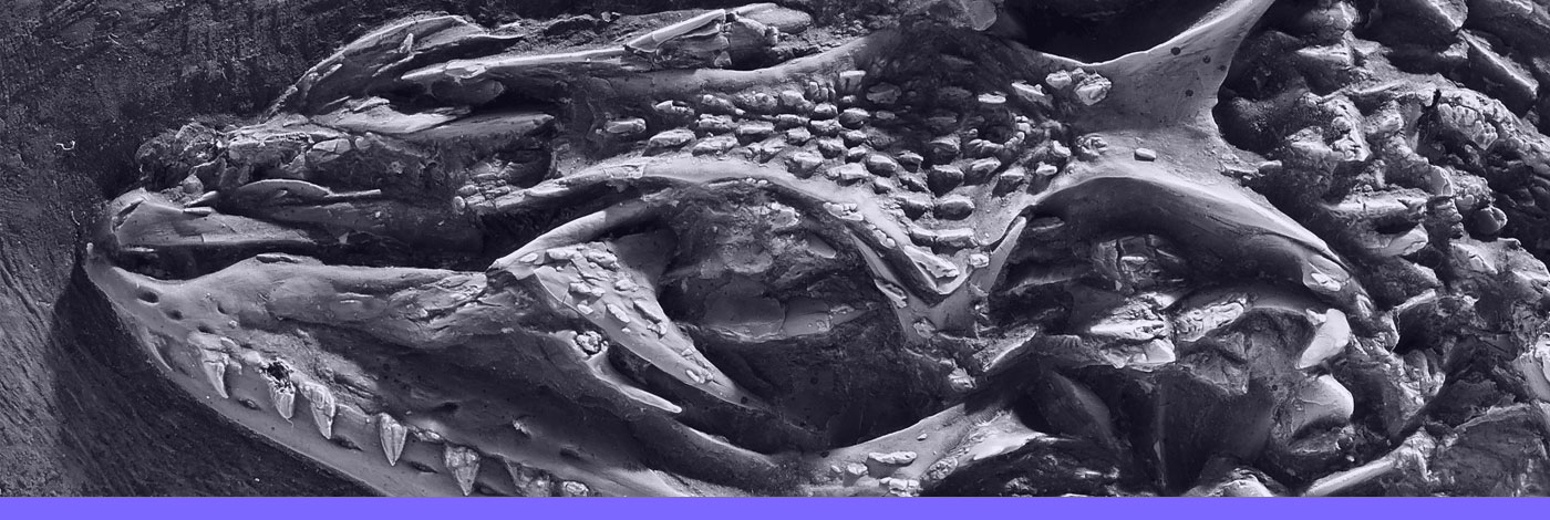
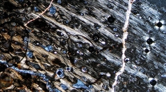
22 (32) - Pages 635-665Data on turtle limb bone histology and microstructure are spotty, especially for Mesozoic taxa, despite significant progress made in recent years. Here we provide first detailed information on the stylopodia of the Late Triassic stem turtles Proganochelys quenstedtii Baur, 1887 from Switzerland and Proterochersis porebensis Szczygielski & Sulej, 2016 from Poland. In both taxa we observed large, internal medullary regions filled with endosteal trabeculae and poorly to moderately vascularized parallel-fibered (grading locally to lamellar) periosteal cortices. Primary vasculature is predominantly longitudinal, in Proterochersis porebensis locally with radial inclination. In large specimens, secondary remodeling is significant in the deeper cortex, but it neither completely obliterates the primary tissue nor reaches the external surface of the bone in either taxon. Comparison of histological data, limb morphology, shell and limb lengths as well as proportions reveal differences in growth patterns between the taxa: Proganochelys quenstedtii seems to grow faster during early life stages than Proterochersis porebensis and attained distinctly larger body sizes earlier in ontogeny, even though the asymptotic body size is roughly the same for both species. Overall, the histological and microstructural characteristics of stylopodial bones of Triassic turtles more closely resemble those of more recent representatives of that group than earlier stem turtles.
Humerus, femur, histology, microstructure, Proterochersis, Proganochelys, turtle, Testudinata, Triassic, Norian