 Comptes Rendus Palevol
15 (1-2) - Pages 176-196
Comptes Rendus Palevol
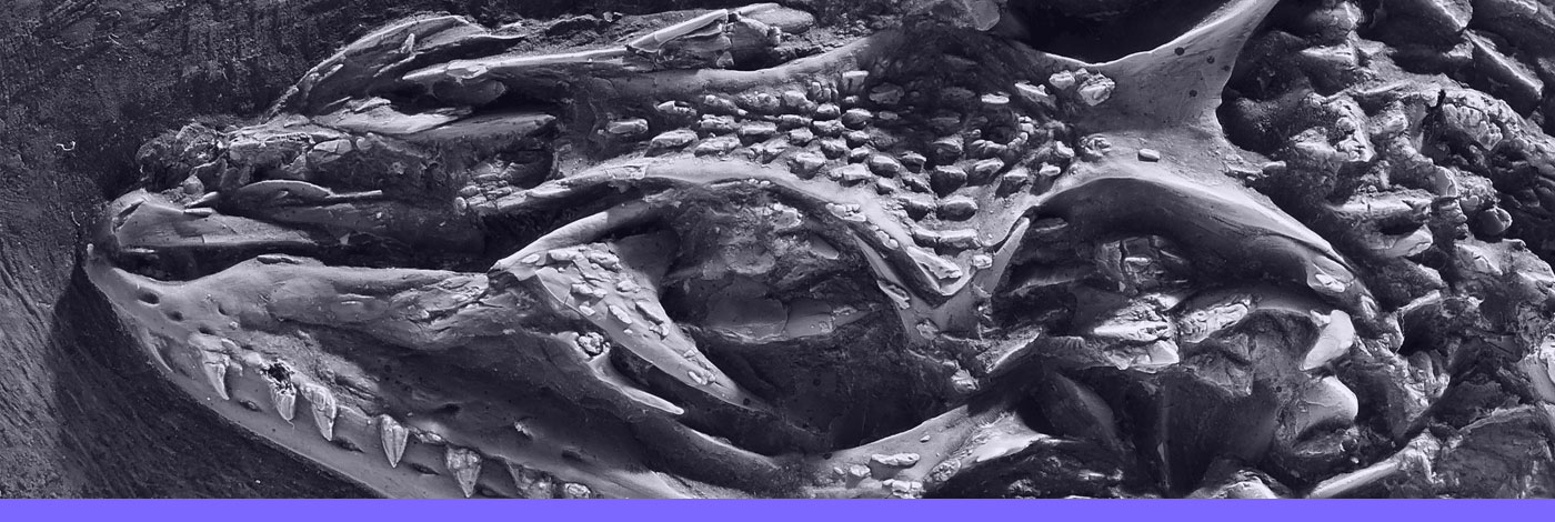
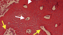
15 (1-2) - Pages 176-196Evolutionary biologists define “metaplasia” as the permanent transformation of a cell identity, and there are many examples of such transformations in living vertebrates (e.g., chondrocytes transforming directly into osteoblasts). These metaplasias have been observed during the mineralization of “ossified” tendons of living birds. In the present study, we examined “ossified” tendons in Bubo and Meleagris and used the characteristics of these metaplastic tissues to recognize them in several non-avian dinosaur taxa. The fossilized skeletal elements that form our sample are varied and include hadrosaurian tendons and a nasal bone, an ankylosaur tail club “handle”, sauropod neural spines, and some dromaeosaur tail rods. The extant avian mineralized tendons were formed of a primary tissue (analogous to primary bone) and secondary reconstructions (SRs; analogous to secondary osteons). Both were composed of fiber bundles (or fascicles) that were closely packed together and separated by arc-shaped spaces in cross-section. When viewed longitudinally, they were arranged in a herringbone pattern. There is no evidence of osteocytes within the primary tendon matrix; what was previously interpreted as osteocyte lacunae are instead arc-shaped spaces between fiber fascicles, and tissue immediately surrounding vascular spaces is dense, avascular and apparently hypermineralized. Mineralization of fibers began centrally and moved in a centrifugal direction. In the non-avian dinosaurs examined, primary and secondary tissue structures were virtually identical to those found within the avian mineralized tendons. Indeed, (1) they were densely fibrous; (2) they showed fiber fascicles separated by arcuate-shaped spaces when viewed transversally, and (3) they were arranged in a herringbone pattern longitudinally. SRs differ from typical Haversian systems in possessing highly irregular borders, suggesting destruction of the fibrous matrix and formation of initial vascular spaces was accomplished perhaps by phagocytosis or enzymatic lysis with subsequent remodeling by fibrocytes, fibroblasts, or an as of yet unknown cell type. Osteocytes with canaliculi were only observed in “mature” SRs, found deep within the elements (and never close to their external borders). Because fossilized primary and secondary tissue structures were identical to those found within the avian mineralized tendons examined, it is likely that identical processes are responsible for their formation. Biomechanical properties were also likely similar, potentially affording carbon fiber-like, trauma-resistant properties to the “ossified” tendons and nasal bones of hadrosaurs, the tail “handles” of ankylosaurs, and the tail rods of dromaeosaurs. In contrast, the primary tissue from a sauropod mineralized nuchal ligament appears to be made of hypermineralized fibrocartilage, but the SRs interdigitating with the hypermineralized fibrocartilage resemble the reconstructions observed in the other fossil skeletal elements and likely formed by the same processes. Since no osteocyte lacunae were observed in any of these dinosaurian primary tissues, we hypothesize that the fossilized cranial and skeletal elements examined here formed through metaplastic transformation (perhaps from fibroblasts) rather than by periosteal and intramembranous ossification. This study suggests that alternative modes of mineralization might be more abundant in non-avian dinosaurs than previously reported.
Metaplasia, “Ossified” tendons, Dinosauria, Ornithischia, Saurischia, Aves, Alternative mode of “ossification”
DOI (Digital Object Identifier)
https://doi.org/10.1016/j.crpv.2015.01.006
Permalink (to link this page)
http://cr-palevol.fr/15/17