 Comptes Rendus Palevol
12 (3) - Pages 149-158
Comptes Rendus Palevol
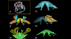
12 (3) - Pages 149-158Barremian and Cenomanian rhynchonelliform brachiopods, one Santonian and one Holocene species (Rhynchonellida and Terebratulida), have been observed using X-ray Computed Tomography in order to investigate the shell interior. Compared to transverse serial sections used formerly, X-ray CT is a promising tool. It permits 3D modelling of the brachidium, which is important for brachiopod classification. Four types of brachidium present during the Cretaceous have been observed (crura for the Rhynchonellida, short loop for Terebratulidina–Terebratulidae, ring-loop for Terebratulidina–Cancellothyrididae and long-loop for Terebratellidina), as well as relationships lophophore/brachidium in a Holocene species. Limitations are discussed, including the nature of the sediments enclosed between the valves, diagenesis, and the necessity to observe several age groups concerning the Terebratellidina. Finally, this non-destructive tool facilitates 3D reconstruction of inaccessible brachidia to improve brachiopod taxonomy and as an aid in curating collections.
Brachiopod, X-ray CT, Brachidium, 3D imagery, Taxonomy, Diagenesis, Collections