 Comptes Rendus Palevol
10 (5-6) - Pages 357-366
Comptes Rendus Palevol
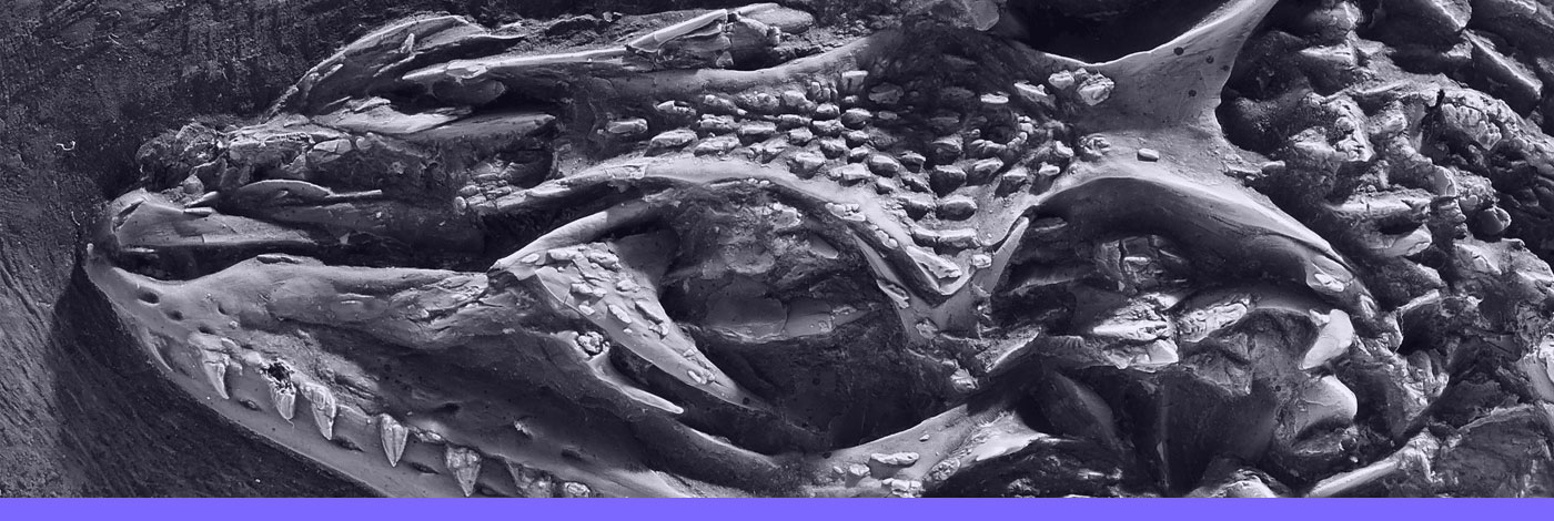
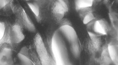
10 (5-6) - Pages 357-366A transmission electron microscope (TEM) study was initiated on samples of geological ages ranging from Devonian to Jurassic to analyse the ultrastructure of the organic matrix in fossil bones that have preserved a histological structure after demineralisation. All samples show a network of variably well-preserved fibrils. Within the sampling, the best results were obtained in two specimens: the scales of the Devonian sarcopterygian tetrapodomorph Eustenopteron foordi, and the humerus of Jurassic dinosaur Lappentosaurus madagascariensis. Despite an extended time difference between both specimens, their fossil bone is composed of a plywood-like structure in which the fibrils are very closely packed. These observations support the hypothesis that dense initial packing of collagen fibrils favours the preservation of the fossil bone.
Collagen, Fossil bone, Vertebrates, TEM, Palaeohistology