
 Cryptogamie, Bryologie
32 (3) - Pages 211-220
Cryptogamie, Bryologie
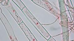
32 (3) - Pages 211-220The aim of this study is to develop a technique of visualization and staining of nuclei of protonemal cells of Ceratodon purpureus (Hedw.) Brid. in order to establish a test of genotoxicity (micronucleus assay). This technique is part of the development of methodology for environmental risk assessment. Bryophytes are often used as biological models in biomonitoring and ecotoxicology. For ecotoxicology studies, there are bioassays, including micronucleus assay. The genotoxicity assay is often practiced in higher plants; we wanted to expand its use to lower plants, like mosses and especially Ceratodon purpureus. To meet the criteria of micronucleus assay, the cells of protonemata of Ceratodon purpureus were selected as a model. The liquid culture of protonemata provides the quantity of biomass needed to achieve a bioassay. First, the development of a reproducible method for visualization and staining of the nuclei of protonemal cells was a necessary and prior step to the realization of micronucleus assay. So for this staining stage, the coloring agent used was the acetic orcein. It is the reference coloring agent for the micronucleus test on Vicia faba (AFNOR, 2004). With our assays of staining, we could develop a reproducible technique of staining of protonemal nuclei. Then, to attempt to visualize micronuclei, we used our method of staining after an exposure of protonemata to cadmium. The first test of staining of micronuclei following exposure of protonemata to Cd failed to demonstrate micronuclei in the conditions of the experiment. Further research must be conducted using other clastogenic agents to validate our results. Assays on micronuclei of cells of protonemata of Ceratodon purpureus are a first in scientific research. Its innovative aspect encourages further research.