 European Journal of Taxonomy
733 (56) - Pages 56-71
European Journal of Taxonomy
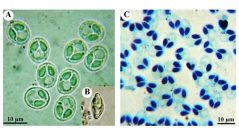
733 (56) - Pages 56-71We report a new myxozoan, Myxobolus opsaridiumi sp. nov., infecting the ornamental fish Opsaridium ubangiensis (Pellegrin, 1901) collected from the Anga River near the city of Yaounde, Cameroon. Plasmodia were found in the skin, muscles and spleen. The overall prevalence of infection was 54.7% (288 parasitized fish out of 526 examined). The myxospores were ovoid to subspherical in frontal view and lenticular in lateral view. The valves were symmetrical and relatively thick, without edge markings. The myxospore measurements were 10.7 ± 0.14 (10–11.5) μm long, 9 ± 0.15 (8–10) μm wide and 6.2 ± 0.7 (5.6–7.2) μm thick. The two ovoid polar capsules were equal in size, converging and opening together at the anterior end, measuring 5 ± 0.07 (4.3–6.0) μm long and 2.7 ± 0.07 (2.2–3.0) μm wide. Polar filaments were coiled from 5 to 7 turns. Histopathological analysis revealed no inflammatory reaction associated with the infection. A BLAST search found that the newly obtained 18 rDNA sequence had a low sequence similarity with available sequences for Myxobolus on GenBank. A phylogenetical analysis based on ribosomal DNA partial sequences showed that M. opsaridiumi sp. nov. is closely associated with several species of Myxobolus infecting cyprinid fish.