 Cryptogamie, Algologie
29 (4) - Pages 285-291
Cryptogamie, Algologie
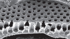
29 (4) - Pages 285-291A technique for obtaining cross-sections of diatom frustules which retains the integrity of the frustule without recourse to an ultra-microtome and allows their examination under SEM is described. The technique employs the polishing and etching of resin-embedded material and can be applied to cleaned diatoms from field samples or cultures. Examples are shown of the types of frustule ultrastructure information that can be obtained using this technique.