 Geodiversitas
29 (1) - Pages 143-216
Geodiversitas
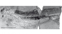
29 (1) - Pages 143-216The anatomy of the Upper Devonian jawless vertebrate Euphanerops longaevus Woodward, 1900, from the Escuminac Formation of Miguasha, Quebec, Canada, is described on the basis of new specimens, some of which display what is regarded here as an extensively mineralized endoskeleton that is essentially made of calcium phosphate, with traces of diagenetic calcite and silicate. Most of the mineralized elements of E. longaevus display the same spongiose microstructure, which notably occurs in such undoubtedly endoskeletal elements, as the fin radials, and this suggests that they all are actually endoskeletal elements. Their structure consists of groups of large, generally paired, ovoid spaces that recall the chondrocytes of lamprey cartilage, and are therefore referred to as “chondrocyte spaces”. The latter are surrounded by a shell of “mineralized territorial matrix”, and cemented by a finely spherulitic “mineralized interterritorial matrix” that extends between them. The question whether this mineralized tissue is an unusual form of biogenic calcified cartilage, or an authigenic, microbially induced post-mortem phosphatization, is discussed, but no definite answer is proposed. The snout of E. longaevus displays three “head stains” that may be either the imprints of the collapsed median olfactory organ and paired eyes, or traces of cartilaginous plates arming the snout. In large specimens, these are followed posteriorly by a large patch of mineralized tissue tentatively interpreted as a braincase. The branchial apparatus consists of an elongated, cone-shaped “basket”, composed of at least 30 vertical, sinuous gill arches, and extending from beneath the presumed braincase to the anal region. The gill arches bear a large number of more or less horizontal gill ray-like mineralized rods, which probably supported the gill filaments. The gill arches seem to have been attached dorsally and ventrally to series of massive endoskeletal elements, referred to as the “copular elements”. The ventral series of copular elements is prolonged anteriorly by a median “anterior ventral rod”, which extends to a ring-shaped structure, the “annular cartilage”. The homology of the latter to the annular cartilage of lampreys remains uncertain. The position of the heart remains problematical, despite the possible presence of a pericardiac cartilage at the rear of the branchial basket. The viscera were housed dorsal to the branchial apparatus, and comprised a large stomach containing fine-grained sediment, but the organization of the digestive tract and its relations to the branchial apparatus remains unknown in detail. The axial skeleton clearly displays complete dorsal and ventral series of arcualia, ventrally to which extends a series of elements referred to as “haemal series”. The anal fin radials are supported by large “anal fin supports”. E. longaevus is regarded here as having possessed thin paired fin radials, arranged in ventrolateral series, which diverge anteriorly, like in anaspids, but this species is unique among vertebrates in having paired fins that extend ventrolaterally to the branchial apparatus. Many anatomical features of E. longaevus remain nevertheless unexplained, such as the “white line” and the “black lines”, tentatively interpreted here as blood vessels. Peculiar mineralized elements, referred to as the “intermuscular elements” and “diffuse mineralized matter”, have no equivalent in other vertebrates and may either have been endoskeletal elements housed in intermuscular connective tissues of the trunk, or haphazardly distributed authigenically phosphatised soft tissues. The oblique, elongated imprints, variously referred to as “scales”, or “myomere imprints” in previous descriptions, are only seen in the smaller and poorly mineralized individuals but their nature remains unknown. The sediment in the stomach contents rather suggests microphagous bottom feeding. The Late Devonian Endeiolepis aneri Stensiö, 1939 (a probable junior synonym of E. longaevus) and the Middle Devonian Achanarella trewini Newman, 2002 and Cornovichthys blaauweni Newman & Trewin, 2001 share with E. longaevus the same organization of the three “head stains” and the same structure and elongation of the branchial apparatus. These four taxa are thus likely to form a clade, the Euphaneropidae. The Lower Silurian Jamoytius kerwoodi White, 1946, may also turn out to belong to this clade. Owing to the uncertainty as to the biogenic or diagenetic nature of the anatomical features described in E. longaevus, no character analysis is proposed. Only a few possible homologies are uniquely shared by euphaneropids and either lampreys or anaspids, or both.
Vertebrata, Euphaneropidae, Devonian, taphonomy, anatomy, homologies